Showing posts with label Thoracic Trauma. Show all posts

Thursday, February 5, 2015

Thoracic Trauma - Traumatic Cardiac Tamponade


Cardiac tamponade is a clinical syndrome caused by the accumulation of fluid in the pericardial space /sac (Sac surrounding the heart), resulting in reduced ventricular filling and subsequent hemodynamic compromise. It can occur from both a medical and traumatic etiology.  In this post we will discuss Traumatic Cardiac Tamponade. The condition is a traumatic emergency, the complications of which include pulmonary edema, shock, and death.

The speed in which the fluid builds up in the pericardial sac is the largest factor in survival.  The faster the fluid accumulates the higher the mortality.  This is why traumatic cardiac tamponade are often more dangerous than those of a medical cause. Rapid accumulation of as little as 150mL of fluid can result in a marked increase in pericardial pressure and can severely impede cardiac output,[2] whereas 1000 mL of fluid may accumulate over a longer period without any significant effect on diastolic filling of the heart. This is due to adaptive stretching of the pericardium over time. A more compliant pericardium can allow considerable fluid accumulation over a longer period without hemodynamic insult.

Heart with pericardial sac opened.


X-ray showing the heart surrounded by a fluid filled pericardia sac.


Signs and symptoms

Symptoms vary with the acuteness and underlying cause of the tamponade. Patients with acute tamponade may present with dyspnea, tachycardia, and tachypnea. Cold and clammy extremities from hypoperfusion are also observed in some patients. Other symptoms may include the following:
  • Elevated jugular venous pressure (JVD)
  • Pulsus paradoxus
 
Beck triad

Described in 1935 by Claude Beck, this complex of physical findings, also called the acute compression triad, refers to increased jugular venous pressure, hypotension, and diminished heart sounds. These findings result from a rapid accumulation of pericardial fluid. This classic triad is usually observed in patients with acute cardiac tamponade.
 
The concept was developed by Claude Beck, a resident and later Professor of Cardiovascular Surgery at Case Western Reserve University.[


Management

Prehospital treatment.

The prehospital treatment of pericardial tamponade is mainly supportive.  Position of comfort if
thermodynamically stable, semifowlers in in respiratory distress, supine if in hypoperfusion,.  High concentration O2, temperature maintenance, rapid transport to the appropriate facility (trauma center).

In Hospital treatment

Removal of pericardial fluid is the definitive therapy for tamponade and can be done using the following three methods:
  • Emergency subxiphoid percutaneous drainage
  • Echocardiographically guided pericardiocentesis
  • Percutaneous balloon pericardiotomy
The role of medication therapy in cardiac tamponade is limited.


Pericardiocentesis


Wednesday, February 4, 2015

Thoracic Trauma - Flail Segment and Pulmonary Contusions


Flail Segment


A flail chest occurs when a segment of the thoracic (rib) cage is separated from the rest of the chest wall. This is usually defined as at least two fractures per rib (producing a free segment), in at least two ribs. A segment of the chest wall that is flail is unable to contribute to lung expansion. Large flail segments will involve a much greater proportion of the chest wall and may extend bilaterally or involve the sternum. In these cases the disruption of normal pulmonary mechanics may be large enough to require mechanical ventilation
 
.The main significance of a flail chest however is that it indicates the presence of an underlying pulmonary contusion. In most cases it is the severity and extent of the lung injury that determines the care need and the possible requirement for mechanical ventilation. Thus the management of flail chest consists of standard management of the rib fractures and of the pulmonary tusions underneath.
 
Diagnosis
Most significant chest wall injuries will be identified by physical examination. Bruising, grazes or seat-belt signs are visible on inspection, and palpation may reveal the crepitus associated with broken ribs. Awake patients will complain of pain on palpation of the chest wall or on inspiration.
A flail chest is identified as paradoxical movement of a segment of the chest wall - ie indrawing on inspiration and moving outwards on expiration. This is often better noted by palpation than by inspection.
 
Pulmonary Contusion


 A pulmonary contusion is an injury to lung tissue, leading to edema and blood collecting in alveolar spaces and loss of normal lung structure & function. This blunt lung injury develops over the course of 24 hours, leading to poor gas exchange, increased pulmonary vascular resistance and decreased lung compliance. There is also a significant inflammatory reaction to blood components in the lung, and 50-60% of patients with significant pulmonary contusions will develop bilateral Acute Respiratory Distress Syndrome (ARDS).
 
Acute respiratory distress syndrome (ARDS) is a life-threatening lung condition that prevents enough oxygen from getting to the lungs and into the blood. Infants can also have respiratory distress syndrome.

Causes

ARDS can be caused by any major direct or indirect injury to the lung. Common causes include:
  • Breathing vomit into the lungs (aspiration)
  • Inhaling chemicals
  • Lung transplant
  • Pneumonia
  • Septic shock (infection throughout the body)
  • Trauma
ARDS leads to a buildup of fluid in the air sacs (alveoli). This fluid prevents enough oxygen from passing into the bloodstream.
The fluid buildup also makes the lungs heavy and stiff, which decreases the lungs' ability to expand. The level of oxygen in the blood can stay dangerously low, even if the person receives oxygen from a ventilator through a endotracheal tube.

 
Pulmonary contusions occur in approximately 20% of blunt trauma patients and it is the most common chest injury in children. The reported mortality ranges from 10 to 25%, and 40-60% of patients will require mechanical ventilation. The complications of pulmonary contusion are ARDS, as mentioned, and respiratory failure, atelectasis.
 
(Atelectasis (at-uh-LEK-tuh-sis) is a condition in which one or more areas of your alveoli collapse or don't inflate properly. If only a small area or a few small areas of the alveoli are affected, you may have no signs or symptoms.
If a large area or several large areas of the alveoli are affected, they may not be able to deliver enough oxygen to your blood.)
 
Diagnosis
 
Pulmonary contusions are rarely diagnosed on physical examination. The mechanism of injury may suggest blunt chest trauma, and there may be obvious signs of chest wall trauma such as bruising, rib fractures or flail chest. These suggest the presence of an underlying pulmonary contusion. Crackles may be heard on auscultation but are rarely heard in the emergency room and are non-specific.
Severe bilateral pulmonary contusions may present with hypoxia - but more usually hypoxia develops as the pulmonary contusions blossom or as a result of subsequent ARDS.
 
In hospital radiological testing (plain chest x-ray or CAT scan) is best way to diagnose a pulmonary contusion. 

Monday, February 2, 2015

Thoracic Trauma - Tension Pneumothorax

 
Thoracic Trauma Pneumothorax - Tension
 
Tension Pneumothorax
 
Tension pneumothorax is the progressive build-up of air within the pleural space, usually due to a lung laceration which allows air to escape into the pleural space but not to return. Positive pressure ventilation may exacerbate (worsen) this 'one-way-valve' effect.
 
Progressive build-up of pressure in the pleural space pushes the mediastinum to the opposite hemithorax, and obstructs venous return (Inferior and superior vena cava)  to the heart. This leads to circulatory instability and may result in traumatic arrest. The classic signs of a tension pneumothorax are deviation of the trachea away from the side with the tension, a hyper-expanded chest, an increased percussion note and a hyper-expanded chest that moves little with respiration.
 
However these classic signs are usually absent and more commonly the patient is tachycardic and tachypneic, and may be hypoxic. These signs are followed by circulatory collapse with hypotension and subsequent traumatic arrest with pulseless electrical activity (PEA). Breath sounds  may be very difficult to interpret and misleading in the high noise environment of the field. There may also be "referred' sounds from the uninjured side that further complicates diagnosis.
 
Tension pneumothorax may develop insidiously (slowly, without obvious symptoms at first, so that the person is not aware of it developing), especially in patients (COPD, trauma. Asthma)  with positive pressure ventilation (BVM / CPAP). This may happen immediately or some hours down the line. An unexplained tachycardia, hypotension and rise in airway pressure are strongly suggestive of
a developing tension pneumothorax.
 
 
Left side tension pneumothorax.  Note that heat is displaced to patients right side.  Also trachea is shifted to right (away from side with pneumothorax).


EMS Treatment of a Tension Pneumothorax
 
Note:  The information in this section are general guidelines and should be consider permission or instruction in actual patient care.  Follow your local guidelines and medical control in all situations.
 
 
  • BLS treatment
    • O2, rapid transport to trauma center
    • If open (sucking) chest wound consider sealing on three side with occlusive dressing or using commercial sealing device.
    • If patient's condition deteriorates after sealing with occlusive dressing remove dressing and see if patient's condition improves.
    • Avoid positive pressure ventilation if at all possible.
  • ALS treatment
    • All of the above
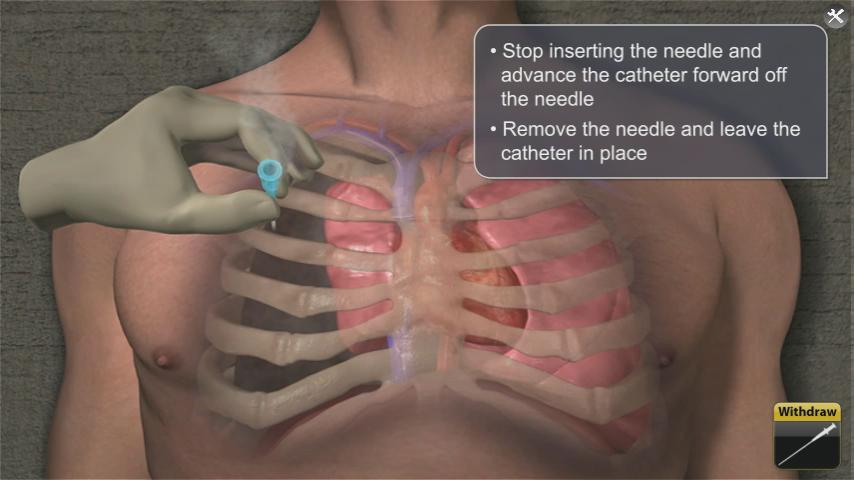
    • Consider needle Thoracostomy over affected lung to relieve pressure
    • 2nd intercostal space (Between 2nd and 3rd rib). Insert needle superior to 3rd rib to avoid neuro-vascular bundle under 2nd rid.
    • Continue to monitor patient.
 
 
Open or sucking chest wound
 
 
Occlusive dressing
 
 
Asherman chest seal
 
 
Bolin Chest seal


SAM chest seal
 
 
Needle Thoracostomy (ALS Level)


Needle Thoracostomy land marking (2nd intercostal space)