Friday, February 6, 2015

First Responders: New Square Hatzolah - EMS, Ebola Training

 
New Square - Earlier this night gathered up to 40 EMT's and Paramedics to the New Square Hatzolah Garage on Reagan Rd. To get important information how to treat a Ebola patient.
The course was given by the Captain Training/Quality Assurance Coordinator Frank Deschino
from Rockland Paramedics Services
Photos: YossiK/First Responders


Thursday, February 5, 2015

Thoracic Trauma - Traumatic Cardiac Tamponade


Cardiac tamponade is a clinical syndrome caused by the accumulation of fluid in the pericardial space /sac (Sac surrounding the heart), resulting in reduced ventricular filling and subsequent hemodynamic compromise. It can occur from both a medical and traumatic etiology.  In this post we will discuss Traumatic Cardiac Tamponade. The condition is a traumatic emergency, the complications of which include pulmonary edema, shock, and death.

The speed in which the fluid builds up in the pericardial sac is the largest factor in survival.  The faster the fluid accumulates the higher the mortality.  This is why traumatic cardiac tamponade are often more dangerous than those of a medical cause. Rapid accumulation of as little as 150mL of fluid can result in a marked increase in pericardial pressure and can severely impede cardiac output,[2] whereas 1000 mL of fluid may accumulate over a longer period without any significant effect on diastolic filling of the heart. This is due to adaptive stretching of the pericardium over time. A more compliant pericardium can allow considerable fluid accumulation over a longer period without hemodynamic insult.

Heart with pericardial sac opened.


X-ray showing the heart surrounded by a fluid filled pericardia sac.


Signs and symptoms

Symptoms vary with the acuteness and underlying cause of the tamponade. Patients with acute tamponade may present with dyspnea, tachycardia, and tachypnea. Cold and clammy extremities from hypoperfusion are also observed in some patients. Other symptoms may include the following:
  • Elevated jugular venous pressure (JVD)
  • Pulsus paradoxus
 
Beck triad

Described in 1935 by Claude Beck, this complex of physical findings, also called the acute compression triad, refers to increased jugular venous pressure, hypotension, and diminished heart sounds. These findings result from a rapid accumulation of pericardial fluid. This classic triad is usually observed in patients with acute cardiac tamponade.
 
The concept was developed by Claude Beck, a resident and later Professor of Cardiovascular Surgery at Case Western Reserve University.[


Management

Prehospital treatment.

The prehospital treatment of pericardial tamponade is mainly supportive.  Position of comfort if
thermodynamically stable, semifowlers in in respiratory distress, supine if in hypoperfusion,.  High concentration O2, temperature maintenance, rapid transport to the appropriate facility (trauma center).

In Hospital treatment

Removal of pericardial fluid is the definitive therapy for tamponade and can be done using the following three methods:
  • Emergency subxiphoid percutaneous drainage
  • Echocardiographically guided pericardiocentesis
  • Percutaneous balloon pericardiotomy
The role of medication therapy in cardiac tamponade is limited.


Pericardiocentesis


Measles (Rubeola)

Measles (Rubeola)


We have by now all heard about the measles outbreak that started in Disneyland or Disney California Adventure Park in Anaheim, California in December, 2014.  To date there have been over 100 cases of measles linked to this out break.  cases have shown up in 14 states including New York.
below is some information about measles. 

Measles is a highly contagious respiratory disease caused by a virus. It spreads through the air through coughing and sneezing. Measles starts with a fever, runny nose, cough, red eyes, and sore throat, and is followed by a rash that spreads all over the body. About three out of 10 people who get measles will develop one or more complications including pneumonia, ear infections, or diarrhea. Complications are more common in adults and young children.

Signs and Symptoms

The symptoms of measles generally appear about seven to 14 days after a person is infected.
Measles typically begins with
  • high fever,
  • cough,
  • runny nose (coryza), and
  • red, watery eyes (conjunctivitis).
 
Two or three days after symptoms begin, tiny white spots (Koplik spots) may appear inside the
mouth.

Koplik Spots ( tiny white spots in the mouth)


Mouth of a patient with Koplik spots, an early sign of measles infection.

Three to five days after symptoms begin, a rash breaks out. It usually begins as flat red spots that appear on the face at the hairline and spread downward to the neck, trunk, arms, legs, and feet. Small raised bumps may also appear on top of the flat red spots. The spots may become joined together as they spread from the head to the rest of the body. When the rash appears, a person’s fever may spike to more than 104° Fahrenheit.
After a few days, the fever subsides and the rash fades.


Measles Rash

Skin of a patient after 3 days of measles infection.

Image of measles infection


Transmission of Measles


boy sneezingMeasles is a highly contagious virus that lives in the nose and throat mucus of an infected person. It can spread to others through coughing and sneezing. Also, measles virus can live for up to two hours on a surface or in an airspace where the infected person coughed or sneezed. If other people breathe the contaminated air or touch the infected surface, then touch their eyes, noses, or mouths, they can become infected. Measles is so contagious that if one person has it, 90% of the people close to that person who are not immune will also become infected.
Infected people can spread measles to others from four days before to four days after the rash appears.

Measles is a disease of humans; measles virus is not spread by any other animal species.


Complications

Measles can be a serious in all age groups. However, children younger than 5 years of age and adults older than 20 years of age are more likely to suffer from measles complications.

Common Complications

Common measles complications include ear infections and diarrhea.
  • Ear infections occur in about one out of every 10 children with measles and can result in permanent hearing loss.
  • Diarrhea is reported in less than one out of 10 people with measles.

Severe Complications

Some people may suffer from severe complications, such as pneumonia (infection of the lungs) and encephalitis (swelling of the brain). They may need to be hospitalized and could die.
  • As many as one out of every 20 children with measles gets pneumonia, the most common cause of death from measles in young children.
  • About one child out of every 1,000 who get measles will develop encephalitis (swelling of the brain) that can lead to convulsions and can leave the child deaf or mentally retarded.
  • For every 1,000 children who get measles, one or two will die from it.
Measles may cause pregnant woman to give birth prematurely, or have a low-birth-weight baby.

Frequently Asked Questions about Measles in the U.S.


Q: Has measles been eliminated from the United States?

A: Yes. In 2000, the United States declared that measles was eliminated from this country. The United States was able to eliminate measles because it has a highly effective measles vaccine, a strong vaccination program that achieves high vaccine coverage in children and a strong public health system for detecting and responding to measles cases and outbreaks.


Q: What does "measles elimination" mean?

A: Measles elimination is defined as the absence of continuous disease transmission for 12 months or more in a specific geographic area. Measles is no longer endemic (constantly present) in the United States.


Q: If measles is eliminated, why do people still get it in the United States?

A: Every year, measles is brought into the United States by unvaccinated travelers (Americans or foreign visitors) who get measles while they are in other countries. They can spread measles to other people who are not protected against measles, which sometimes leads to outbreaks. This can occur in communities with unvaccinated people.
Most people in the United States are protected against measles through vaccination, so measles cases in the U.S. are uncommon compared to the number of cases before a vaccine was available. Since 2000, when measles was declared eliminated from the U.S., the annual number of people reported to have measles ranged from a low of 37 people in 2004 to a high of 644 people in 2014.


Q: Where do cases of measles that are brought into the United States come from?

A: Measles can be brought into the United States from any country where the disease still occurs or where outbreaks are occurring including Europe, Africa, Asia, and the Pacific. In recent years, many measles cases have been brought into the United States from common U.S. travel destinations, such as England, France, Germany, India, and, during 2014, from the Philippines and Vietnam.
 

Q: Why have there been more measles cases in the United States in recent years?

A: In 2008, 2011, 2013 and 2014, there were more reported measles cases compared with previous years. CDC experts attribute this to:
  • more measles cases than usual in some countries to which Americans often travel (such as England, France, Germany, India, the Philippines and Vietnam), and therefore more measles cases coming into the US, and/or
  • more spreading of measles in U.S. communities with pockets of unvaccinated people.
 

Q: How effective is the measles vaccine?

A: The measles vaccine is very effective. One dose of measles vaccine is about 93% effective at preventing measles if exposed to the virus and two doses is about 97% effective.


Q: Could I still get measles if I am fully vaccinated?

A: Very few people—about three out of 100—who get two doses of measles vaccine will still get measles if exposed to the virus. Experts aren’t sure why; it could be that their immune systems didn’t respond as well as they should have to the vaccine. But the good news is, fully vaccinated people who get measles are much more likely to have a milder illness, and they are also less likely to spread the disease to other people, including people who can’t get vaccinated because they are too young or have weakened immune systems.


Q: Do I ever need a booster vaccine?

A: No. People who received two doses of measles vaccine as children according to the U.S. vaccination schedule are considered protected for life and do not ever need a booster dose.
Adults need at least one dose of measles vaccine, unless they have evidence of immunity. Adults who are going to be in a setting that poses a high risk for measles transmission, including students at post-high school education institutions, healthcare personnel, and international travelers, should make sure they have had two doses separated by at least 28 days.
If you’re not sure whether you were vaccinated, talk with your doctor.

Q: Am I protected against measles?

A: You are considered protected from measles if you have written documentation (records) showing at least one of the following:
  • You received two doses of measles-containing vaccine, and you are a(n)—
    • school-aged child (grades K-12)
    • adult who was not vaccinated as a child and will be in a setting that poses a high risk for measles transmission, including students at post-high school education institutions, healthcare personnel, and international travelers.
  • You received one dose of measles-containing vaccine, and you are a(n)—
    • preschool-aged child
    • adult who was not vaccinated as a child and will not be in a high-risk setting for measles transmission.
  • A laboratory confirmed that you had measles at some point in your life.
  • A laboratory confirmed that you are immune to measles.
  • You were born before 1957.


Q: What should I do if I’m unsure whether I’m immune to measles?

A: If you’re unsure whether you’re immune to measles, you should first try to find your vaccination records or documentation of measles immunity. If you do not have written documentation of measles immunity, you should get vaccinated with measles-mumps-rubella (MMR) vaccine. Another option is to have a doctor test your blood to determine whether you’re immune, but this option is likely to cost more and will take two doctor’s visits. There is no harm in getting another dose of MMR vaccine if you may already be immune to measles (or mumps or rubella).

Q: How common was measles in the United States before the vaccine?

A: Before the measles vaccination program started in 1963, we estimate that about 3 to 4 million people got measles each year in the United States. Of those people, 400 to 500 died, 48,000 were hospitalized, and 4,000 developed encephalitis (brain swelling) from measles.


Q: Is measles a concern for the United States?

A: Yes. Since measles is still common in many countries, this disease will continue to be brought into the United States. Measles is highly contagious, so anyone who is not protected against measles is at risk of getting the disease. People who are unvaccinated for any reason, including those who refuse vaccination, risk getting infected with measles and spreading it to others, including those who cannot get vaccinated because they are too young or have specific health conditions.


Q: Could measles ever re-establish itself in the United States?

A: Yes, it is possible that measles could become endemic (constant presence of a disease in an area) in the United States again, especially if vaccine coverage levels drop. This can happen when people
  • forget to get vaccinated on time,
  • don’t know that they need a vaccine dose (this is most common among adults), or
  • refuse vaccines for religious, philosophical or personal reasons.
Research shows that people who refuse vaccines tend to group together in communities. When measles gets into communities with pockets of unvaccinated people, outbreaks are more likely to occur. These communities make it difficult to control the spread of the disease and make us vulnerable to having the virus re-establish itself in our country.
High sustained measles vaccine coverage and rapid public health response are critical for preventing and controlling measles cases and outbreaks.


Q: Will the United States ever get rid of measles completely?

A: Yes, it's possible. The first step is to eliminate measles from each country and region of the world. Once this happens, there will be no place from which measles can spread.
All member states in the six World Health Organization regions have committed to eliminating measles by the year 2020. Once a disease has been eliminated from every country, it is considered "eradicated" from the world. See the Measles and Rubella Initiative for more information.


Wednesday, February 4, 2015

Thoracic Trauma - Flail Segment and Pulmonary Contusions


Flail Segment


A flail chest occurs when a segment of the thoracic (rib) cage is separated from the rest of the chest wall. This is usually defined as at least two fractures per rib (producing a free segment), in at least two ribs. A segment of the chest wall that is flail is unable to contribute to lung expansion. Large flail segments will involve a much greater proportion of the chest wall and may extend bilaterally or involve the sternum. In these cases the disruption of normal pulmonary mechanics may be large enough to require mechanical ventilation
 
.The main significance of a flail chest however is that it indicates the presence of an underlying pulmonary contusion. In most cases it is the severity and extent of the lung injury that determines the care need and the possible requirement for mechanical ventilation. Thus the management of flail chest consists of standard management of the rib fractures and of the pulmonary tusions underneath.
 
Diagnosis
Most significant chest wall injuries will be identified by physical examination. Bruising, grazes or seat-belt signs are visible on inspection, and palpation may reveal the crepitus associated with broken ribs. Awake patients will complain of pain on palpation of the chest wall or on inspiration.
A flail chest is identified as paradoxical movement of a segment of the chest wall - ie indrawing on inspiration and moving outwards on expiration. This is often better noted by palpation than by inspection.
 
Pulmonary Contusion


 A pulmonary contusion is an injury to lung tissue, leading to edema and blood collecting in alveolar spaces and loss of normal lung structure & function. This blunt lung injury develops over the course of 24 hours, leading to poor gas exchange, increased pulmonary vascular resistance and decreased lung compliance. There is also a significant inflammatory reaction to blood components in the lung, and 50-60% of patients with significant pulmonary contusions will develop bilateral Acute Respiratory Distress Syndrome (ARDS).
 
Acute respiratory distress syndrome (ARDS) is a life-threatening lung condition that prevents enough oxygen from getting to the lungs and into the blood. Infants can also have respiratory distress syndrome.

Causes

ARDS can be caused by any major direct or indirect injury to the lung. Common causes include:
  • Breathing vomit into the lungs (aspiration)
  • Inhaling chemicals
  • Lung transplant
  • Pneumonia
  • Septic shock (infection throughout the body)
  • Trauma
ARDS leads to a buildup of fluid in the air sacs (alveoli). This fluid prevents enough oxygen from passing into the bloodstream.
The fluid buildup also makes the lungs heavy and stiff, which decreases the lungs' ability to expand. The level of oxygen in the blood can stay dangerously low, even if the person receives oxygen from a ventilator through a endotracheal tube.

 
Pulmonary contusions occur in approximately 20% of blunt trauma patients and it is the most common chest injury in children. The reported mortality ranges from 10 to 25%, and 40-60% of patients will require mechanical ventilation. The complications of pulmonary contusion are ARDS, as mentioned, and respiratory failure, atelectasis.
 
(Atelectasis (at-uh-LEK-tuh-sis) is a condition in which one or more areas of your alveoli collapse or don't inflate properly. If only a small area or a few small areas of the alveoli are affected, you may have no signs or symptoms.
If a large area or several large areas of the alveoli are affected, they may not be able to deliver enough oxygen to your blood.)
 
Diagnosis
 
Pulmonary contusions are rarely diagnosed on physical examination. The mechanism of injury may suggest blunt chest trauma, and there may be obvious signs of chest wall trauma such as bruising, rib fractures or flail chest. These suggest the presence of an underlying pulmonary contusion. Crackles may be heard on auscultation but are rarely heard in the emergency room and are non-specific.
Severe bilateral pulmonary contusions may present with hypoxia - but more usually hypoxia develops as the pulmonary contusions blossom or as a result of subsequent ARDS.
 
In hospital radiological testing (plain chest x-ray or CAT scan) is best way to diagnose a pulmonary contusion. 

Monday, February 2, 2015

CPAP for NYS EMTs is coming

                                                                  CPAP FOR EMS

Continuous Positive Airway Pressure (CPAP) devices will be coming to NYS EMT's shortly.  Here is a short guide to CPAP.  Since we do not know yet how the NYS BLS Protocols will permit EMTs to use CPAP we will keep it very general for now. It will obviously be used in Acute Pulmonary Edema, but it is unclear if it will be allowed in other emergency situation.

Before we start just remember that CPAP cannot be used in any patient with any of the following conditions EVEN if they are in respiratory distress:

1. Decreased Mental Status / Inability to sit up / Altered Mental Status
2. Respiratory arrest / Apnea /  Respiratory failure / poor inspiratory effort
3. Pneumothorax / Trauma to the thorax / Subcutaneous Emphysema
4. Any form of Shock / Hypotension
5. Nausea / Vomiting / any risk of aspiration
6. Facial Trauma / Abnormalities


How CPAP Works:

Continuous Positive Airway Pressure, (CPAP), is the maintenance of positive pressure throughout the complete respiratory cycle, (inspiration and expiration), when breathing spontaneously. CPAP is not the same as Positive End Expiratory Pressure or PEEP. PEEP only provides pressure on the expiratory side by offering resistance to exhalation using a spring-loaded valve or air flow.

 
1. During the inspiratory phase, patients in distress will have to create a higher flow rate of air to meet their needs, this will present as an increase in work-of-breathing (WOB). Patients in respiratory distress may need to achieve inspiratory flow rates greater than 65 liters per min (LPM). With the inspiratory support of CPAP, the patient does not have to work as hard to inhale and overcoming the auto PEEP in the lung. Auto-PEEP represents the abnormal, and usually undetected, residual pressure above atmospheric remaining in the alveoli at end-exhalation due to air trapping.  The auto PEEP has to be overcome each time a person inhales, for a healthy person the work is minimal and goes unnoticed. However, those patients with stiff lungs, (CHF and Pulmonary Fibrosis), have to work extremely hard to overcome the increased auto PEEP on every breath.
 
2. The increased inspiratory pressure also increases the size, therefore the surface area of the
alveoli, providing a greater opportunity for gas exchange or respiration. The process increases the
Functional Residual Capacity (FRC) of the lung. The FRC is the area where gas exchange takes place.
 
3. Since a greater oxygen percentage is able to reach the alveoli during CPAP, the partial pressure
of the oxygen molecule will be greater. The higher partial pressure will allow more oxygen to
diffuse into the blood stream improving oxygenation.
 
4. Fluid in the alveolar space cannot only make the lung stiff, (increasing inspiratory
work-of-breathing), it also creates a barrier that can reduce gas exchange. The pressure from CPAP
can reduce the fluid by forcing fluid out of the alveolar space back into the interstitium.
 
5. During the expiratory phase, the patient will breathe against a threshold of resistance that
works as a pneumatic splint to hold the airways open. Patients with chronic lung disease have
weakened airways that have a tendency to collapse on expiration, causing air trapping. Having the
airways stinted open during exhalation will make inspiration on the next breath less difficult.
 
6. The resistance during exhalation can open non-ventilated areas of the lung recruiting alveoli
that have collapsed due to atelectasis, (a collapse of lung tissue affecting part, or all, of one
lung effecting gas exchange).
 7. CPAP decreases pre-load and after-load on the heart reducing the heart’s workload. However, a reduction in pre-load and after-load will have an effect on the patient’s blood pressure. Patients
should have a systolic blood pressure of at least 100 mmHg before starting CPAP.
 
What Types of Patients Are NOT Candidates for CPAP?
Being able to assess and determine who is, and who is not, a candidate for CPAP has a great impact on whether CPAP will be effective or not. CPAP can be a very effective treatment for patients in respiratory distress but is not indicated for patients in respiratory failure. Respiratory distress patients are still compensating even though they may be working hard. It is not uncommon for respiratory distress patients to have oxygen saturations (SpO2 ) and carbon dioxide, (CO2), levels within normal range. Key determinants include; is the patient alert, (even though they are working hard), and can they follow directions. Patients that have gone into respiratory failure may exhibit a decrease in work-of-breathing, CO2 levels climbing, oxygen saturations falling, and their level of consciousness declining, (most likely from CO2 narcosis). CPAP is not indicated for respiratory failure patients.


CPAP and Congestive Heart Failure (CHF):
The treatment of Congestive Heart Failure, (CHF), by EMS has changed significantly in the last couple of years. The mainstays of CHF treatment (ALS) in the pre-hospital setting are CPAP and nitroglycerin. The efficacy of Lasix and Morphine is under scrutiny and has been removed from many EMS ALS protocols. Many systems that adopt CPAP start with CHF. The effects of CPAP are well suited for the physiologic issues associated with this disease. Pulmonary edema, associated with CHF, makes the lungs stiff and it is difficult for the patient to inhale; and can be observed as difficulty breathing during the inspiratory phase of ventilation or inspiratory shortness-of-breath. Since CHF is primarily a heart problem, CPAP addresses the side effects of a failing heart and its impact on the lungs.  CHF is a process that will continue to spiral down the cardiogenic shock pathway until the cycle is broken. As the patient’s heart fails, more fluid ends up in the lungs. With more fluid in the lungs, less oxygen makes it to the heart muscle, so the heart fails even more. The first step in stopping the cycle is the early use of CPAP. If the patient does not have lung disease then the airways should function normally and not collapse on exhalation. In this case, the inspiratory pressure will force the fluid out of the lungs, expand the alveoli which, in turn, will increase gas exchange (respirations) improving oxygenation.In addition, with the airways being held open by the expiratory resistance, the patient does not have to overcome
the auto PEEP at the beginning of each breath. With the improvement of gas exchange and reduced impact of auto PEEP, the patient’s work-of-breathing will be reduced. With the reduced work, there will be less stress on the heart. A key factor in the initiation of CPAP is the patient’s blood pressure. It is recommended that the systolic blood pressure be at least 100 mmHg before starting CPAP due to the reduction in pre-load and after-load.
 
More to come after the protocols are released.


Thoracic Trauma - Tension Pneumothorax

 
Thoracic Trauma Pneumothorax - Tension
 
Tension Pneumothorax
 
Tension pneumothorax is the progressive build-up of air within the pleural space, usually due to a lung laceration which allows air to escape into the pleural space but not to return. Positive pressure ventilation may exacerbate (worsen) this 'one-way-valve' effect.
 
Progressive build-up of pressure in the pleural space pushes the mediastinum to the opposite hemithorax, and obstructs venous return (Inferior and superior vena cava)  to the heart. This leads to circulatory instability and may result in traumatic arrest. The classic signs of a tension pneumothorax are deviation of the trachea away from the side with the tension, a hyper-expanded chest, an increased percussion note and a hyper-expanded chest that moves little with respiration.
 
However these classic signs are usually absent and more commonly the patient is tachycardic and tachypneic, and may be hypoxic. These signs are followed by circulatory collapse with hypotension and subsequent traumatic arrest with pulseless electrical activity (PEA). Breath sounds  may be very difficult to interpret and misleading in the high noise environment of the field. There may also be "referred' sounds from the uninjured side that further complicates diagnosis.
 
Tension pneumothorax may develop insidiously (slowly, without obvious symptoms at first, so that the person is not aware of it developing), especially in patients (COPD, trauma. Asthma)  with positive pressure ventilation (BVM / CPAP). This may happen immediately or some hours down the line. An unexplained tachycardia, hypotension and rise in airway pressure are strongly suggestive of
a developing tension pneumothorax.
 
 
Left side tension pneumothorax.  Note that heat is displaced to patients right side.  Also trachea is shifted to right (away from side with pneumothorax).


EMS Treatment of a Tension Pneumothorax
 
Note:  The information in this section are general guidelines and should be consider permission or instruction in actual patient care.  Follow your local guidelines and medical control in all situations.
 
 
  • BLS treatment
    • O2, rapid transport to trauma center
    • If open (sucking) chest wound consider sealing on three side with occlusive dressing or using commercial sealing device.
    • If patient's condition deteriorates after sealing with occlusive dressing remove dressing and see if patient's condition improves.
    • Avoid positive pressure ventilation if at all possible.
  • ALS treatment
    • All of the above
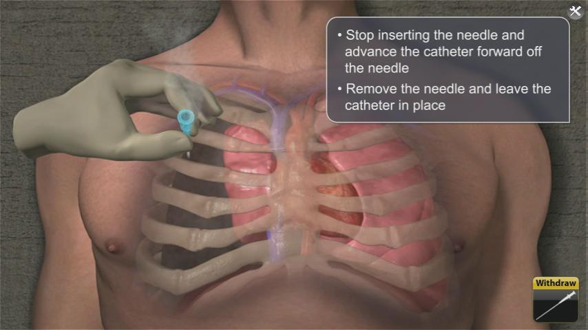
    • Consider needle Thoracostomy over affected lung to relieve pressure
    • 2nd intercostal space (Between 2nd and 3rd rib). Insert needle superior to 3rd rib to avoid neuro-vascular bundle under 2nd rid.
    • Continue to monitor patient.
 
 
Open or sucking chest wound
 
 
Occlusive dressing
 
 
Asherman chest seal
 
 
Bolin Chest seal


SAM chest seal
 
 
Needle Thoracostomy (ALS Level)


Needle Thoracostomy land marking (2nd intercostal space)


Friday, June 27, 2014

EMS Myth -There's no such thing as too much oxygen. What we should be saying: - Can Oxygen Hurt???

Disclaimer - This post contains information that may be in direct contradiction with our NYS BLS protocols.  I am not advocating that any NYS certified EMS provider violate any part of our outdated protocols.

                               EMS

EMTs in NYS have been instructed by our protocols and some well-meaning Instructors (both not up to date with current research) that patients should receive high concentration oxygen via non rebreathing oxygen masks in every situation.  That is unless the patient is medically savvy enough to refuse O2. 
A quick review of current medical research reveals that this theory has been under attack for years.  It is now widely recognized that oxygen like every other substance in our body needs to be maintained within a normal range.  Too little (hypoxia) is bad, but too much (hyperoxia) may be worse. The AHA in its current ACLS guidelines defines a normal O2 saturation as 92 to 94%.  Patients falling in those ranges are considered to be well oxygenated and do not need to be given supplemental oxygen. 
Some people may argue that we should not trust a machine or that most of the times our transport times are so short (Rockland County) that it does not matter.  I would counter by sayings that if the patient was a member of your family would you want them to receive a treatment that may be harmful.  Remember my favorite quote.  "Primum non nocere (not that I can pronounce it)  a Latin phrase that means "first, do no harm."

Read the article below and let’s see what everyone thinks.  

Have a great weekend.  

Frank
*****************************************

Several Studies Point Out Harm in Hyperoxia

Learning Objectives
>> Review current research on supplemental oxygen use in the prehospital field.
>> Evaluate the use of oxygen for myocardial infarction, COPD, stroke and neonatal patients.


Key Terms
Cochrane review: A group of people who prepare reports so that healthcare professionals can make informed decisions about the care they provide.
Cofactor: A substance that acts with another substance to bring about certain effects.
Hyperoxia: A bodily condition characterized by a greater oxygen content of the tissues and organs than normally exists at sea level.
Meta-analysis: A statistical technique for combining the results of several smaller studies into one large study.
Metalloprotein: A conjugated protein in which the prosthetic group is a metal.
Mitochondria: A component of a cell that generates energy for the cell to use.
Odds ratio: A statistical measure of the probability or chance of an event occurring.
Partial pressure: The pressure generated by a gas in a solution; used as a measure of how much gas is present.
Relative risk: A statistical measure of the risk of an event occurring in one group compared with the risk in another group.

Oxygen is a ubiquitous treatment in prehospital care. Most of us remember the excitement of being trained to use it, because it’s typically the first drug that EMS providers are taught to administer. As a core EMS skill, all providers should be highly proficient with oxygen delivery equipment. Less well taught, however, is the fact that oxygen is a drug, which, like all others, has indications, contraindications, adverse effects, a dose and an intended duration of administration.
I remember being taught as a young volunteer EMT to administer oxygen for everything from nausea and vomiting to lower limb fractures. (I was even told at one point that it had analgesic properties.)
This article highlights some current research on supplemental oxygen use in prehospital care and discusses controversies that are arising from this research.
Physiology
Oxygen is used in every cell of the body—except, ironically, red blood cells—to convert chemical potential energy from food into potential energy in the form of adenosine triphosphate (ATP). The mitochondria are the powerhouses of the cells, containing enzymes that gradually break down fats, sugars and proteins into single carbon compounds that release a small amount of energy each time.
At the end of this chain of carbon breakdown, an enzyme called cytochrome oxidase uses oxygen as a cofactor to phosphorylate (i.e., add a phosphorous atom) to adenosine diphosphate (ADP) to produce ATP. ATP is an energy store, allowing the body to use energy when it needs it.
Getting the oxygen to the cells is a bit trickier now than it was when single-celled organisms started using oxygen billions of years ago. A complex circulatory system has evolved to get the oxygen from the alveoli to the cells, and a complex respiratory system has evolved to get oxygen into the blood.
The problem is that oxygen isn’t particularly soluble in blood. So the solution is the hemoglobin contained within the red blood cell. The red blood cells aren’t really cells, because they don’t contain mitochondria, nuclei, Golgi apparatus or all the other things that make cells cells. They’re better thought of as little bags of hemoglobin.
Hemoglobin is a metalloprotein made up of four subunits, each containing a protein (globin) and an iron-containing compound (heme). It’s the iron that’s the key. As we all know, iron and oxygen happily combine to form iron oxide—rust. Rust is red. Oxygenated blood is red. Each time you take a breath in, you cause some of your blood to rust. The beauty of hemoglobin is that it can also “un-rust,” allowing it to offload oxygen to body tissues and be ready to pick more up in the lungs.
The four molecules of hemoglobin work together in a system known as cooperative or allosteric binding. This means that when there isn’t much oxygen around (e.g., in the tissues) hemoglobin doesn’t bind to oxygen very well, so the oxygen dissociates and goes into the tissues. However, when there’s lots of oxygen around (e.g., in the lungs), the hemoglobin avidly binds to oxygen, allowing greater uptake into the blood.
The affinity of hemoglobin for oxygen can also be altered by the conditions of the tissue the blood is flowing through. So in hot, acidotic tissues with high carbon­ dioxide (CO2) levels (e.g., exercising muscle), the oxygen affinity of hemoglobin is poor, allowing more oxygen to be offloaded to the tissues. However, in cold, alkalotic blood with low CO2 levels, the affinity for oxygen is much higher, allowing increased loading in the lungs. This occurs as part of the response to high altitude.
Oxygen directly affects the tissues it travels through by interacting with blood vessels. In most, oxygen acts as a vasoconstrictor, like norepinephrine. It’s thought that this response is part of the cardiovascular system’s function known as autoregulation, whereby organs alter their own blood supply in response to such factors as oxygen and CO2 levels, acidity, potassium and lactate levels in the blood. The exception to this rule is in the lungs, in which a phenomenon known as hypoxic pulmonary vasoconstriction occurs, allowing lung units that are poorly ventilated to autoregulate their blood supply and decrease ventilation perfusion mismatch.
Myocardial Ischemia
Oxygen has been referenced as an integral component of the treatment of myocardial ischemia since 1900. In EMS, it’s considered a primary treatment; indeed the only treatment that can be administered by EMTs or emergency medical responders in many jurisdictions. Hearing that giving supplemental oxygen to patients with cardiac chest pain may not be beneficial—and may even be harmful—may shock many EMS professionals. But the theory isn’t that new.
In 1976, a randomized controlled trial was published in the British Medical Journal in which 157 patients with uncomplicated myocardial infarction (MI) were randomized to receive either supplemental oxygen or air for the first 24 hours following onset of symptoms.1 There was no significant difference in the primary outcome of death or the secondary outcomes of ventricular dysrhythmia or pain requiring analgesia.
There was, however, a non-significant trend toward increased mortality in the group that received oxygen. A caveat of this study is that it took place in the era before revascularization therapy, so care must be taken in comparing it with modern studies of MI management.
Despite this small study, healthcare providers continued to give oxygen for many years. Further, small, randomized studies in the later part of this past century also indicated there may be limited benefit from oxygen use. But it wasn’t until the past year that a systematic review and then a Cochrane review gave a strong indication that oxygen may be harmful in uncomplicated MI.2
The Cochrane review, which analyzed the 1976 paper and two other studies from 1997 and 2004, concluded that the limited evidence available showed no benefit and potential harm from oxygen use. The review indicated that a large, randomized controlled trial should be conducted.
On the basis of the Cochrane review, the American Heart Association (AHA) recommended in the 2010 CPR Guidelines that supplemental oxygen no longer be administered to patients with uncomplicated cardiac chest pain who have an oxygen saturation greater than 94%.3
Cardiac Arrest
A very important paper published in the past year gave a strong indication that hyperoxia may actually be more harmful than hypoxia in adults during and immediately following out-of-hospital cardiac arrest.
A paper in the Journal of the American Medical Association studied the partial pressure of oxygen (PaO2) in arterial blood of patients brought to the emergency department (ED) following successful resuscitation by EMS.4 The patients were stratified based on an arterial blood gas taken within 24 hours of hospital admission into those with hypoxia (PaO2 less than 60 mmHg), those with normoxia (PaO2 61–299 mmHg) and those with hyperoxia (PaO2 greater than 300 mmHg).
The study was multicenter and quite large, enrolling 6,326 patients over five years. The primary outcome was survival to hospital discharge. It found that hyperoxia was a significant independent risk factor for in-hospital mortality. The odds ratio for death was 1.8 (95% CI 1.5–2.2) in the hyperoxia group compared with the normoxia group. This was even higher than the odds ratio for death in the hypoxia group.
The authors of the study correctly note that correlation doesn’t equal causation. However, there’s a plausible mechanism by which hyperoxia could cause increased mortality in patients following cardiac arrest, and the chance of causation is great enough to consider limiting the amount of oxygen given to patients following cardiac arrest to a sufficient amount to keep arterial oxygen saturation 94–96%. The one time 100% oxygen is always indicated (via non-rebreather mask or BVM) for a patient is prior to an intubation, to effectively “wash out” the nitrogen and prevent rapid onset of hypoxia during an intubation attempt.
COPD
Acute exacerbations of chronic obstructive pulmonary disease (COPD) are a common reason to call an ambulance, and supplemental oxygen (often high-flow) is often administered by EMS professionals to these patients. However, a recent randomized controlled trial conducted in the prehospital environment has produced dramatic results indicating that providing high-flow oxygen to patients with an exacerbation of COPD—and possibly even patients with undifferentiated shortness of breath who don’t have a firm diagnosis of COPD—can significantly increase mortality. 5
The study was carried out in Hobart, Australia. Instead of randomizing patients, paramedics were randomized to either provide high-flow oxygen (8–10 LPM via a non-rebreather mask) or titrated oxygen (variable flow via nasal cannula titrated to keep arterial oxygen saturation at 88–92%).5
When nebulized drugs were required, they were delivered by oxygen in the control arm and by compressed air in the intervention arm. The study was extended over a one-year period and randomized 226 patients into the control arm and 179 patients into the intervention arm.
A large number of patients were excluded from both groups after being retrospectively identified as not having a diagnosis of COPD, leaving 117 patients in the control arm and 97 patients in the intervention arm.
Analysis was by intention to treat, and a large number of patients in the intervention arm received high-flow oxygen but were still included in the analysis as though they had received titrated oxygen, making the result of the study all the more remarkable.
The study showed a relative risk of death in the intervention arm of 0.42 (0.02–0.89, p= 0.02) in all patients and a relative risk of death in the intervention arm of 0.22 (0.05–0.91, p=0.04) in those with confirmed COPD. So even in patients without COPD, high-flow oxygen was associated with a doubling of mortality. When the results were analyzed as treatment received rather than intention to treat, the same trends in mortality were identified; however, due to the smaller numbers, the results were no longer statistically significant.
Neonatal Resuscitation
It’s been known for some time that prolonged administration of high oxygen concentrations to premature neonates suffering from apneas is associated with retinopathy of prematurity, which causes blindness. A more recent finding, however, is that resuscitation of neonates with air rather than 100% oxygen results in a decrease in mortality. A 2004 systematic review and meta-analysis pooled the results of five studies involving a total of 1,302 neonates.6
These were all randomized trials of oxygen versus air in the resuscitation of neonates. Three were blinded, and two were unblinded. Although none of the studies showed a significant decrease in mortality, the pooled results showed a significant decrease in mortality with air compared with oxygen. The relative risk of death in the air group was 0.71 (0.54–0.94), with a number needed to harm of 20. (Twenty babies would have to be treated with 100% oxygen to cause one death). The deaths occurred during follow up over a 24-month period. Interestingly, there was no significant increase in such neurological complications as cerebral palsy.
This review has caused a significant change in international protocols for neonatal resuscitation, with the latest edition of the AHA resuscitation Guidelines recommending initial resuscitation of neonates with air, switching only to oxygen when there’s clear evidence of significant hypoxaemia following resuscitation with air.7
Stroke
Modern emergency management of acute stroke has evolved along similar lines to that of myocardial ischemia, with the potential for reperfusion therapy leading to a significant decrease in morbidity and mortality. Supplemental oxygen has long been a standard component of emergency care for patients with acute stroke. However, a study conducted more than a decade ago in Scandinavia cast some doubt on this treatment.8
The study randomized 310 patients with acute ischemic stroke (hemorrhagic stroke and subarachnoid hemorrhage were excluded) to receive either oxygen (3 LPM via nasal cannula) or air for the first 24 hours following hospital admission. Outcomes were survival, measured at one year and stroke severity (using the Scandinavian Stroke Scale) and disability (using the Barthel index)—both measured at 7 months.
The study showed no significant difference in survival (69% in the oxygen group, 73% in the air group, p= 0.3). The authors did, however, comment on a non-significant trend toward decreased survival in the oxygen group.
Analysis of stroke severity and disability index for patients also showed no significant difference, but when a subgroup analysis of patients with severe stroke at presentation was carried out, there was a statistically significant decrease in survival in the oxygen group (82% versus 91%, p=0.023; OR 0.045, 95% CI 0.23–0.9.)
Although these results should not be viewed as conclusive, they suggest oxygen perhaps has no benefit and may be harmful to some patients suffering acute ischemic stroke. On the basis of this study, some EMS services have removed oxygen from the protocols for managing adult patients with uncomplicated acute stroke.
Harm from Supplemental O2
Many theories exist to explain why supplemental oxygen may be harmful. It’s well accepted that prolonged, supranormal blood and tissue oxygen tension is detrimental. Breathing 100% oxygen for longer than three days causes certain death in many species (thankfully, not humans). Breathing 100% oxygen causes thickening of alveolar membranes and restriction of lung expansion. In hyperbaric situations, it can cause seizures.
Hyperoxia also causes free-radical damage. Free radicals are oxygen atoms with a charge due to an unequal number of protons and electrons. These radicals are known to cause intracellular damage and cell destruction (much like pouring hydrogen peroxide into an open would). It’s known that ischemic tissues are particularly sensitive to free-radical damage. This may be a major factor in the pathogenesis of hyperoxia.
Another possibility is that increased oxygen tension causes widespread vasoconstriction. This is known to occur in cerebral arteries, but it may also occur in other vessels, including coronary arteries. This may explain why administration of oxygen following MI may not be beneficial.
Hypoxia—Not So Bad?
In May 2007, a group of intensivists climbed Mt. Everest.9 When they got to the top, they removed their oxygen masks, allowed their bodies time to equilibrate and took arterial blood gas (ABG) samples from each other. The samples were rushed back down to base camp by Sherpa guides and analyzed. The results were startling.
The intent of the study was to see how low the PaO2 could get in the blood of fit and healthy volunteers and then to use this data to guide the management of critically ill patients in intensive care with respiratory failure. The results showed that the average PaO2 was 24.6 mmHg, and the lowest was 19.1 mmHg. The normal PaO2 range for an adult at sea level is 80–100 mmHg. Many clinicians would view the levels achieved in this study as being incompatible with life. The average oxygen saturation (SaO2) was 54.0%, and the lowest SaO2 was 34.4%. These numbers would horrify a paramedic. However, none of these subjects were gravely ill. Some felt giddy and slurred their words slightly. Importantly, no long-term harm came to them. The authors of the paper hope to use their data to further study the limits of hypoxaemia in the critically ill.
Benefits of O2
Having reviewed a few conditions where hyperoxia may be harmful, we must bear in mind that profound and prolonged hypoxia is universally fatal. The timely administration of oxygen can be life saving in many cases. Supplemental oxygen should still be administered to patients who have an oxygen saturation of less than 94% for any reason. (In many jurisdictions, however, it will be acceptable to target an oxygen saturation of greater than 88% in patients with COPD).

Oxygen should also be administered to any patients with actual or potential airway compromise (e.g., epiglottis or airway burns) and drug assists and/or rapid sequence intubations.
The rationale for this treatment isn’t to increase tissue oxygenation but to increase oxygen reserves in the lungs so that if the airway is lost for any reason, the patient will take longer to desaturate before a critical level of hypoxia is reached, which allows providers time to rescue the airway with a supraglottic or surgical airway.
Providers should continue to administer oxygen to patients who have evidence of globally poor tissue perfusion (e.g., hypovolemic or septic shock patients). The rationale for this treatment is to ensure the hemoglobin molecules are fully saturated with oxygen and that there’s as much oxygen as possible dissolved in the blood, increasing the driving pressure to aid internal respiration.
Conclusion
Oxygen is a drug and should be administered to appropriate patients in an appropriate dose. A recent editorial coined the term “Goldilocks effect” to highlight that we should strive to deliver an amount of oxygen that’s not too little, not too much but just right.10 In the 2010 Guidelines, the AHA target an oxygen saturation should be 94%, following successful resuscitation.11
Although we have traditionally erred on the side of caution in prehospital care by providing liberal amounts of supplemental oxygen, we may actually have been erring on the side of harm. EMS providers are skilled professionals who are used to learning the ins and outs of a new drug before administering it appropriately.
Perhaps it’s time we treated oxygen like a new, exciting drug and begin to administer it with the knowledge that, like all other drugs, it has indications, contraindications and, in particular, adverse effects. Until further studies clarify practice and provide a stronger evidence base for guidelines, EMS professionals should at least think before they pick up the mask.
References
1. Rawles J, Kenmure A. Controlled trial of oxygen in uncomplicated myocardial infarction. Br Med J. 1976;1(6018):1121–1123.
2. Cabello J, Burls A, Emparanza J, et al. Oxygen therapy for acute myocardial infarction. Cochrane Database of Syst Rev. 2010;(6):CD007160.
3. O’Connor R, Brady W, Brooks S, et al. Part 10: acute coronary syndromes: 2010 American Heart Association Guidelines for Cardiopulmonary Resuscitation and Emergency Cardiovascular Care. Circulation. 2010;122(18 Suppl 3):S787–817.
4. Kilgannon J, Jones A, Shapiro N, et al. Association between arterial hyperoxia following resuscitation from cardiac arrest and in-hospital mortality. JAMA. 2010;303(21):2165–2171.
5. Austin M, Wills K, Blizzard L, et al. Effect of high flow oxygen on mortality in chronic obstructive pulmonary disease patients in prehospital setting: randomised controlled trial. Br Med J. 2010;341:c5462.
6. Davis P, Tan A, O’Donnell C, et al. Resuscitation of newborn infants with 100% oxygen or air: a systematic review and meta-analysis. Lancet. 2004;364(9442):1329–1333.
7. Kattwinkel J, Perlman J, Aziz K, et al. Part 15: Neonatal Resuscitation: 2010 American Heart Association Guidelines for Cardiopulmonary Resuscitation and Emergency Cardiovascular Care. Circulation. 2010;122(18 Suppl 3):S909–919.
8. Ronning O, Guldvog B. Should stroke victims routinely receive supplemental oxygen: A quasi-randomized controlled trial. Stroke. 1999;30(10):2033–2037.
9. Grocott M, Martin D, Levett D, et al. Arterial blood gases and oxygen content in climbers on Mount Everest. N Engl J Med. 2009;360:140–149.
10. Hommers C. Oxygen therapy post-cardiac arrest: The ‘Goldilocks’ principle? Resuscitation. 2010 (12):1605–1606.
11. Peberdy M, Callaway C, Neumar R, et al. Part 9: post-cardiac arrest care: 2010 American Heart Association Guidelines for Cardiopulmonary Resuscitation and Emergency Cardiovascular Care. Circulation. 2010;122(18 Suppl 3):S768–786.